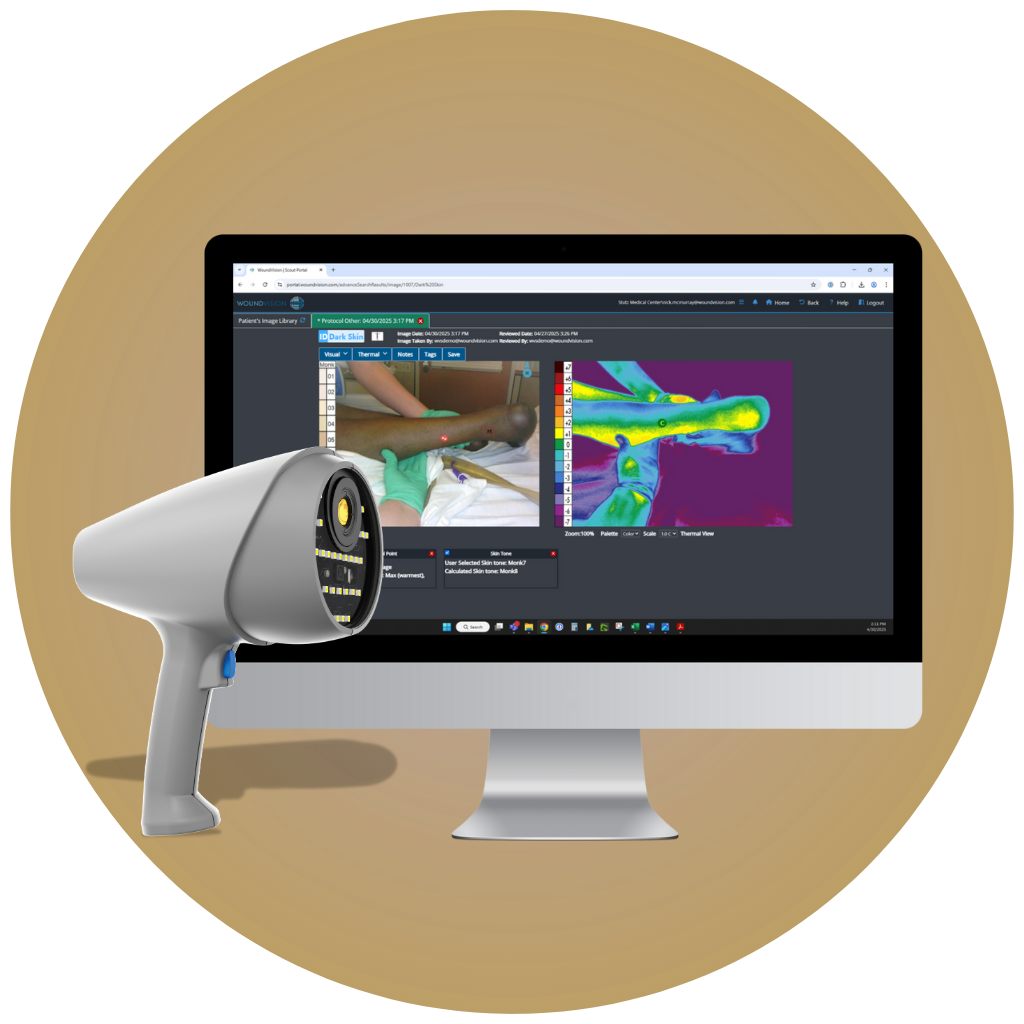
Advancing health equity shouldn't be hard.
We can help.
Announcing integration of the Monk Skin Tone (MST) Scale with the WoundVision Scout software.
Scout + Monk Skin Tone (MST) Scale

As the industry pioneers in dual-modality skin and wound imaging, our foundation has been and always will be long-wave infrared thermography (LWIT). Unlike traditional skin assessments that rely on subjective visual cues—often ineffective across darker skin tones—LWIT is scientifically validated to perform consistently across all patients, regardless of pigmentation.
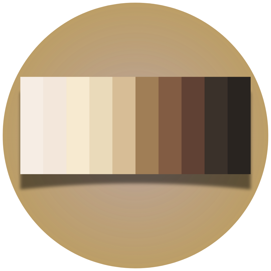
Now, we’re taking the next leap forward by incorporating the Monk Skin Tone (MST) Scale into our Scout software platform, allowing for documentation and analysis of skin tone alongside our thermal imaging data. We believe this fusion of thermal objectivity with skin tone awareness will enable clinicians to better understand patient risk, tailor interventions, and eliminate disparities in documentation.
Check out the information below and schedule a demo to experience the future of skin assessment.

The Monk Skin Tone (MST) Scale is currently available as a research tool only. Contact clinical@woundvision.com to learn more about accessing this powerful feature.
License: Creative Commons Attribution 4.0 International license. Scale Usage Citation: Monk, Ellis. “Monk Skin Tone Scale,” 2019. https://skintone.google.
- The early signs of pressure injury aren’t easily identified in dark skin tones.1
- Pressure injury assessment in dark skin tones is inaccurate and can result in delayed identification.2
- The visual cues associated with DTPI may appear differently in dark skin.1
- Patients with dark skin tones are more likely to develop higher-stage pressure injuries.2
- Discrepancies and systemic biases across healthcare systems can result in patients with dark skin receiving suboptimal care.3
- Studies have shown nursing education lacks diversity in formal training for skin tone assessment.4

The Solution for Assessing Dark Skin Tones
Enhance Your Assessment With Long-Wave Infrared Thermography (LWIT)
Case #1 - Patient with Darkly Pigmented Skin - Left Heel
No Visible Discoloration w/ Relative Skin Temperature Changes
Relative skin temperature changes over areas of ischemia can present as colder than surrounding skin and tissue.¹
Case #2 - Patient with Darkly Pigmented Skin - Right Heel
No Visible Discoloration w/ Relative Skin Temperature Changes
Relative skin temperature changes over areas of inflammation can present as warmer than surrounding skin and tissue.¹
Case #3 - Patient with Darkly Pigmented Skin - Right Ischium
No Visible Discoloration w/ Relative Skin Temperature Changes
Relative skin temperature changes over areas of ischemia can present as colder than surrounding skin and tissue.¹
Case #4 - Patient with Darkly Pigmented Skin - Sacrococcygeal
No Visible Discoloration w/ Relative Skin Temperature Changes
Relative skin temperature changes over areas of inflammation can present as warmer than surrounding skin and tissue.¹
Case #5 - Patient with Darkly Pigmented Skin - Left Heel
No Visible Discoloration w/ Relative Skin Temperature Changes
Relative skin temperature changes over areas of inflammation can present as warmer than surrounding skin and tissue.¹
Case #6 - Patient with Darkly Pigmented Skin - Left Heel
No Visible Discoloration w/ Relative Skin Temperature Changes
Relative skin temperature changes over areas of ischemia can present as colder than surrounding skin and tissue.¹
Power of Temperature
In deep tissue pressure injuries, temperature change often precedes skin color changes, with discoloration appearing differently in dark skin.
Universally Reliable
LWIT is unaffected by skin pigmentation, allowing for universal assessment of patients of all skin tones.
Advocacy
Research has shown LWIT is a powerful, real-time, non-contact, bedside tool that allows clinicians to advocate for patients of all skin tones.
Eliminate Subjectivity
Adding LWIT as an assessment adjunct can improve patient outcomes by providing objective, quantifiable relative temperature data.
Backed by Science, Research and Clinical Practice Guidelines
Retrospective analysis of thermographic imaging of pressure injuries
92.5% sensitivity in identifying pressure injuries on admission across all skin tones.
Statistically outperformed the Braden Scale.
Sustainable Success: Implementing an Infrared Thermography Scanning Protocol in Acute Care
HAPI incidence reduced by 66% per 1,000 patient days.
Estimated return on investment resulted in $1.35MM in avoided penalties and $45K from PSI-03 measure.
Shedding Light on Native American Health Disparities Utilizing Long-Wave Infrared Thermography
“Implementing LWIT resulted in a significant decrease in pressure injury prevalence, the ability to capture and treat vascular wounds and ischemic limbs with quick interventions and providing excellent care for the high population of Native Americans.”
"To date, the facility has captured over 41,000 images on 14,000 patients."
Utilization of Innovation Wound Care Technologies in Pressure Injury Assessment and Treatment: Treatment: A Quality Improvement Wound Ostomy Continence (WOC) Nurse Lead Initiatives
“Long wave infrared technology (LWIT) was added to prevention bundle as adjunct to identify thermal anomalies in Deep Tissue Pressure Injury occurrence, guiding treatment upon early identification.”
“Utilization of LWIT in darker skin tones guided earlier interventions, treatment and assisted with health equity.”
"Estimated $4 to $7 million in savings."
Beyond 4 Eyes in 4 Hours: Implementing Long Wave Infrared Thermography
“HAPI incidence reduced from a baseline of 25.71 (per 1000 discharges) to 2.55.”
“As a result of this project, the 18-month financial gain is estimated at over $11 million dollars.”
Color Blind Clarity - Preventing Pressure Injuries Using Thermography in LTACHs
Utilizing thermography guides the management of pressure injuries in patients with darker pigmented skin tones - improving clinical outcomes.
Clinician and patient education involving thermography increased compliance with the care plan and supported patient participation.
"A Wound Care “View” to Promote Health Equity: Using Long Wave Infrared Thermography (LWIT)
“LWIT immediately visualized unknown health concerns, followed by prompt review and education.”
“The objective was to provide wound ostomy and continence (WOC) nurse assessments and education at a health fair for the inner-city community’s homeless, indigent, and non-English speaking population.”
Ten top tips: assessing darkly pigmented skin
“Consider enhancing your assessment of darkly pigmented skin with technology designed to assess perfusion.”
“Technology to enhance assessments at admission has clear advantages to determining if tissue injury has occurred but is not yet visible.”
Using thermography to assess pressure injuries in patients with dark skin
“Warm tissue is well perfused, cold tissue is not.”
“The visible color of the skin has no impact on the infrared radiation captured by thermography.”


